Research

Dynamic states connect molecular events to cellular decisions.

Biology is often captured as a sequence of static snapshots. We seek to understand the transient states between those snapshots: where they arise, how long they persist, and how they shape cell behaviour.

Scientific premise

The most informative biology often exists between stable endpoints.

Cells continuously reorganise molecular interactions, signalling assemblies, organelles and mechanical structures. These changes can be local, short-lived and heterogeneous, yet they determine how cells sense their environment and choose a response.

Our research asks how such states can be observed directly in living systems and connected to mechanism. Tool development follows from that question, rather than serving as an endpoint of its own.

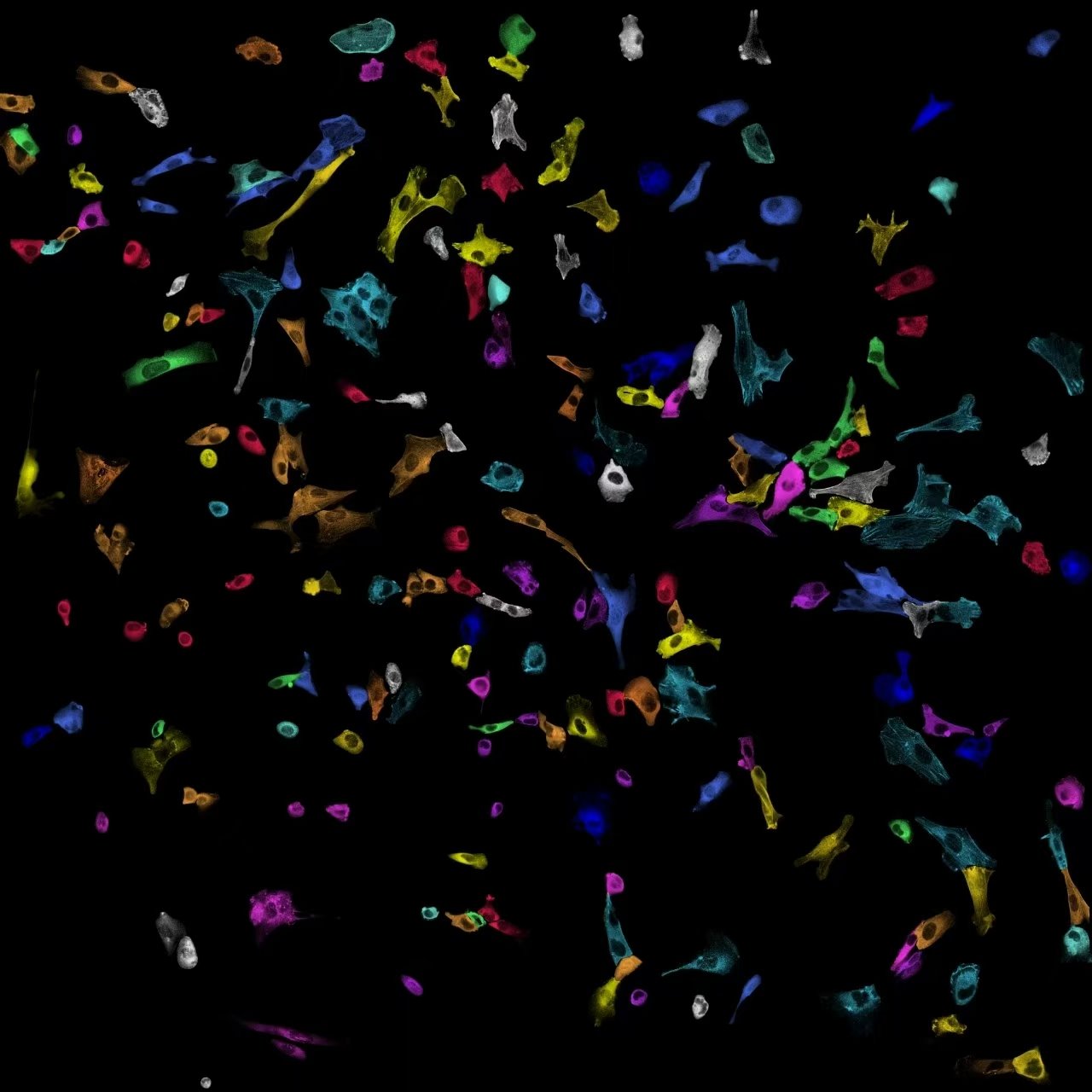
A wide field of individually segmented cells, each rendered in a different colour against a black background
Single cells segmented from one imaging field, each given its own colour.

What we ask

Biological systems where dynamics matter

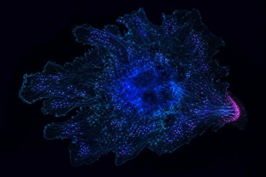
Conceptual visualisation of a macrophage with podosome-scale puncta and a localised immune signalling zone

Immune signalling

How is signalling organised at immune-cell interfaces?

Immune responses emerge from signals assembled and remodelled in specific cellular locations. We investigate how molecular activity is coordinated in macrophages, podosomes and immune synapses, and how local organisation shapes cell behaviour.
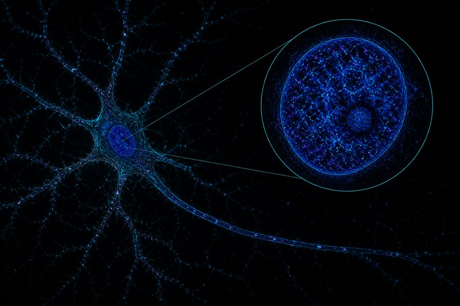
Conceptual visualisation of a neuron with long processes and a magnified view of nuclear speckles, nucleolus and biomolecular condensates

Neuronal nuclear states

How does activity reorganise the neuronal nucleus?

Neuronal activation reaches beyond the membrane and cytoplasm to reshape molecular organisation inside the nucleus. We study how phosphorylation and biomolecular condensation influence nuclear speckles, nucleolar organisation and other dynamic nuclear states.
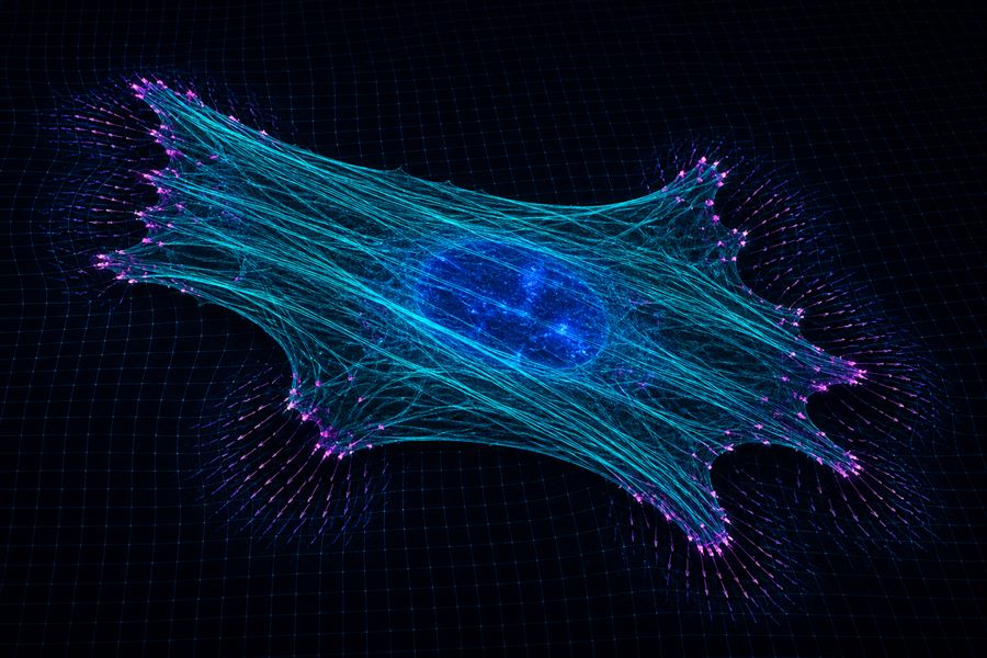
Conceptual visualisation of cellular mechanobiology with stress fibres, focal adhesions, traction forces and substrate deformation

Mechanobiology

How do physical forces become biochemical information?

Cells experience force through adhesion, cytoskeletal tension, membrane deformation and the surrounding matrix. We study how these physical inputs reorganise molecular states and propagate from force-bearing structures toward cellular decisions.
How we answer

Tools for seeing and steering living cells

Each capability addresses a different limit of observation. Their integration enables measurements that are spatially resolved, time-sensitive, molecularly specific and quantitative.

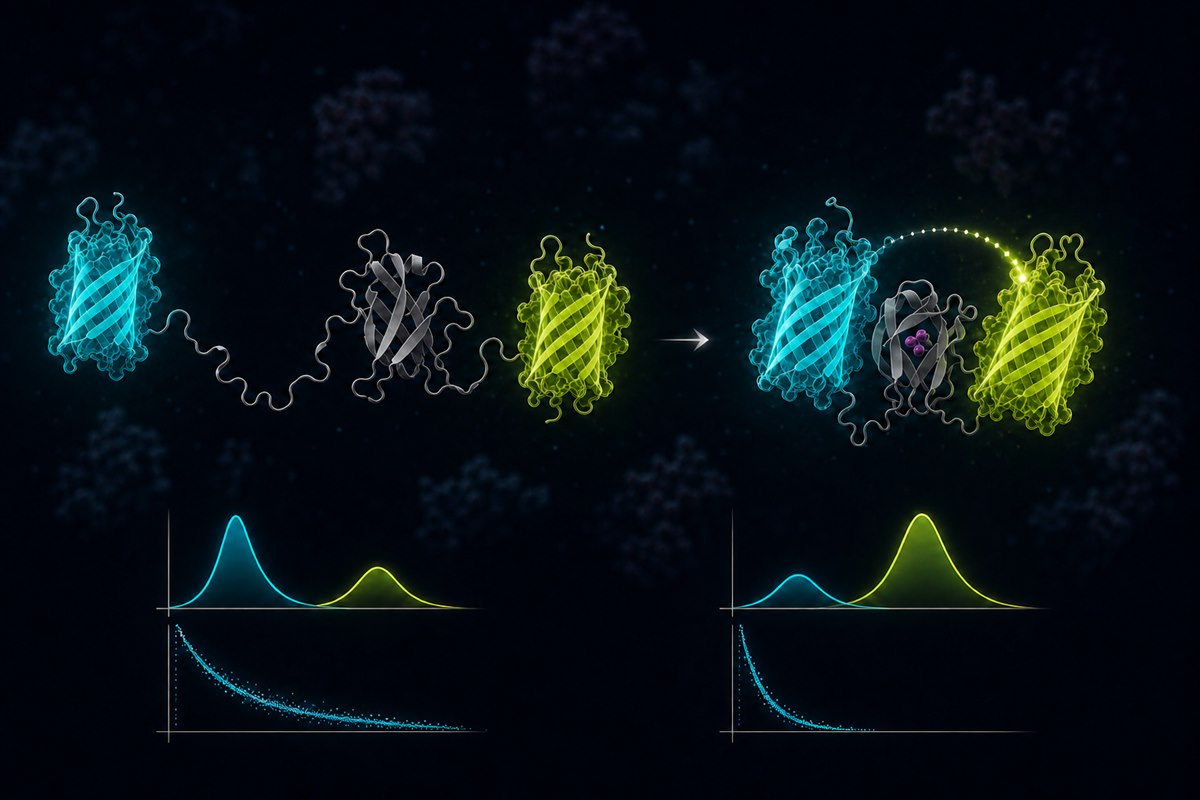
A cyan and a yellow fluorescent protein flanking a sensor domain: when the sensor closes, the two fluorophores come together, energy transfers, and the emission spectrum and fluorescence lifetime both shift

See

Biosensors

Engineered proteins that turn a conformational change into a change in fluorescence — read out by FRET, fluorescence lifetime, or single-molecule tracking in living cells.

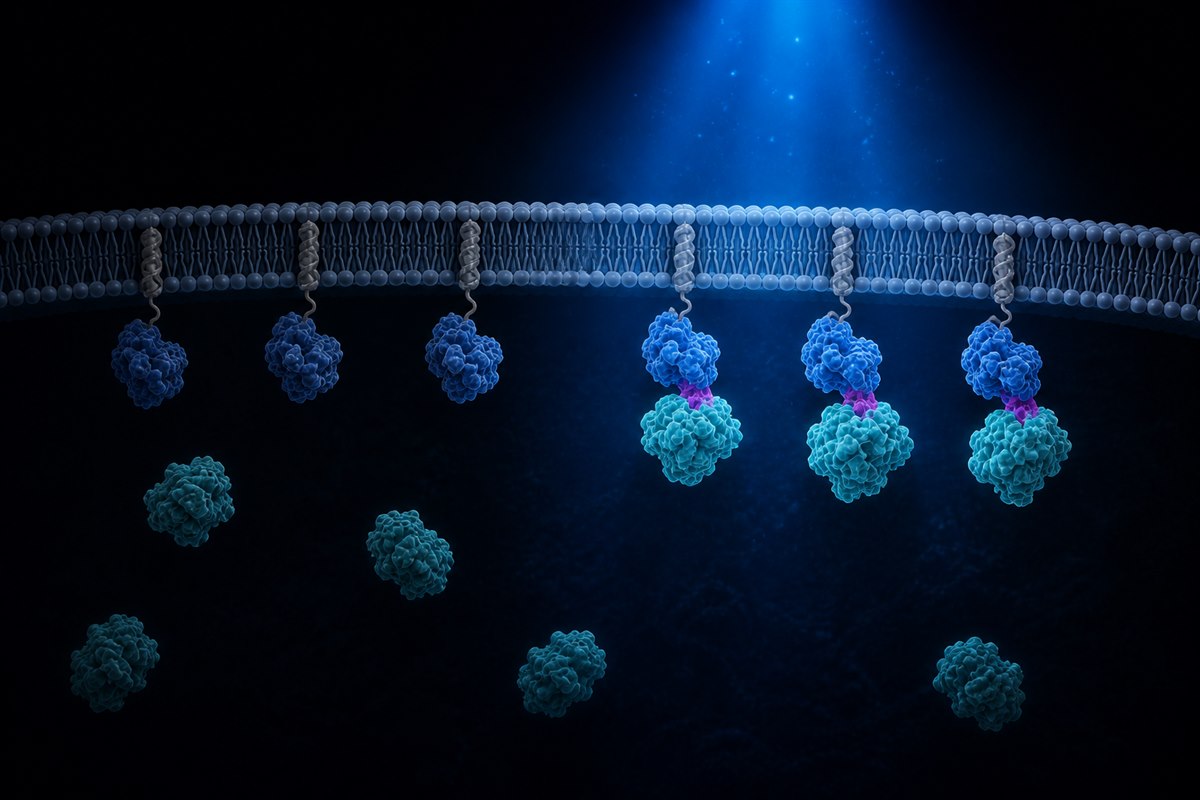
Membrane-anchored proteins under a beam of blue light: the illuminated ones have recruited a partner protein up from the cytoplasm and docked it at the membrane, while those left in the dark remain unbound

Steer

Optogenetics

Light-controlled switches that recruit a protein to a chosen location, or turn its activity on and off — in seconds, and only where we illuminate.

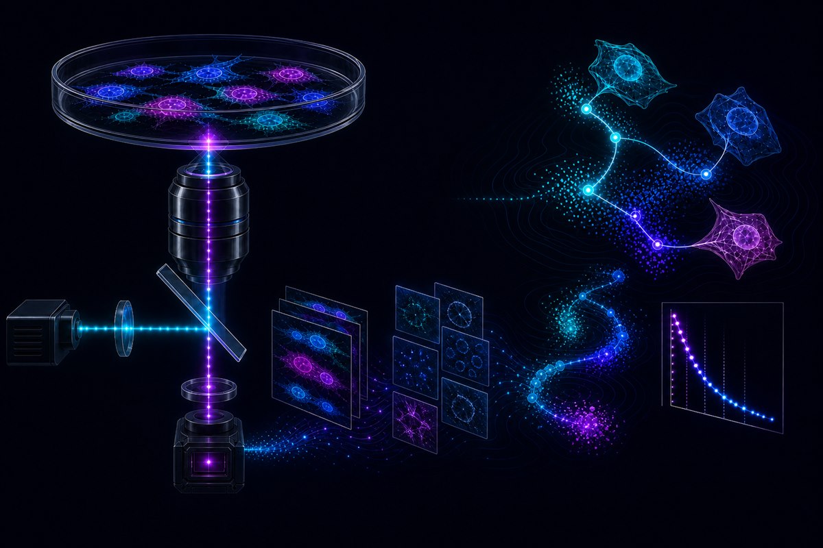
A microscope light path: laser excitation reflects off a dichroic mirror up through the objective into a dish of living cells; the emission returns to the camera, and the recorded image stack is then segmented, tracked from cell to cell, and reduced to a fluorescence-lifetime decay curve

Observe

Microscopy

Imaging systems built in-house for live, quantitative measurement — single-molecule, light-sheet, fluorescence-lifetime and structured illumination — together with the analysis that turns a recording into numbers.

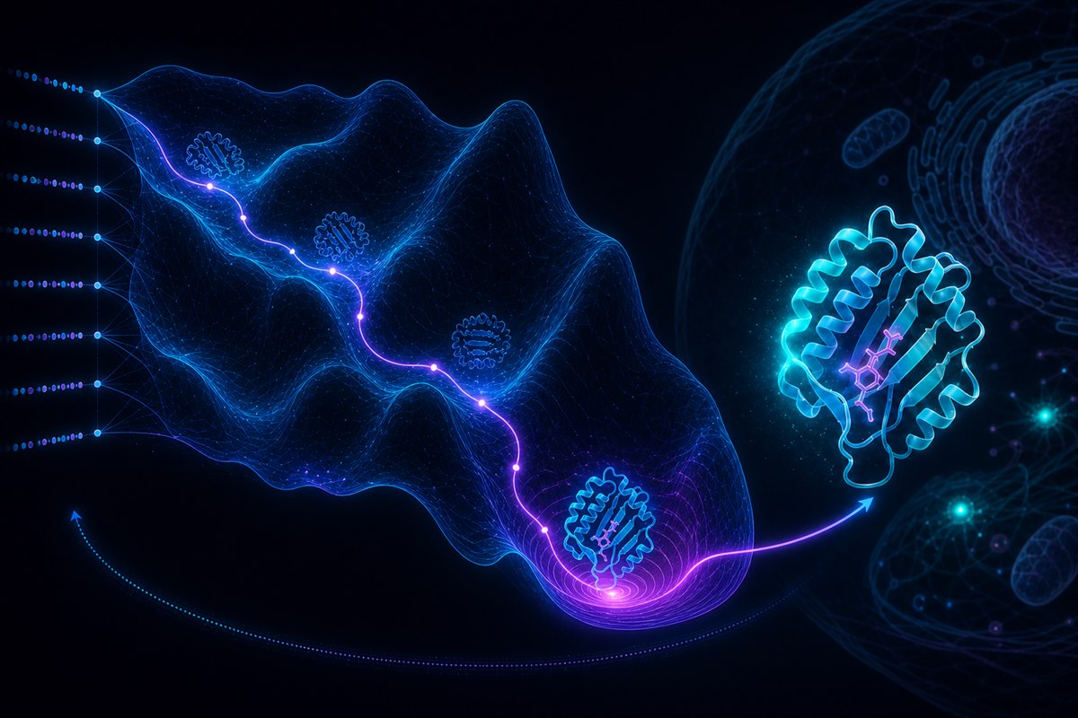
A sequence fed through a neural network traces a path down an energy landscape, passing through successive folds to a minimum, and yields a designed protein that is then tested inside a cell

Design

AI-based protein design

We design biosensors, actuators and binders computationally, then test them directly in living cells — closing the loop between what a measurement requires and what a molecule can do.

How we work

A programme organised around states, not techniques

Observe in living systems

Follow processes as they unfold instead of inferring dynamics from fixed endpoints.

Perturb with precision

Connect observation to mechanism through controlled changes in molecular state, location and timing.

Integrate across scales

Relate molecular events to subcellular organisation, cellular behaviour and system-level responses.

Integrated pipeline

From measurement design to mechanism

The platform is described at the level of scientific capability; specific unpublished implementations remain confidential.

  1. 01Molecular design

    Define the biological state to measure.

  2. 02Advanced imaging

    Observe that state in living systems.

  3. 03AI & computation

    Extract patterns and model transitions.

  4. 04Biological insight

    Test mechanisms in relevant systems.